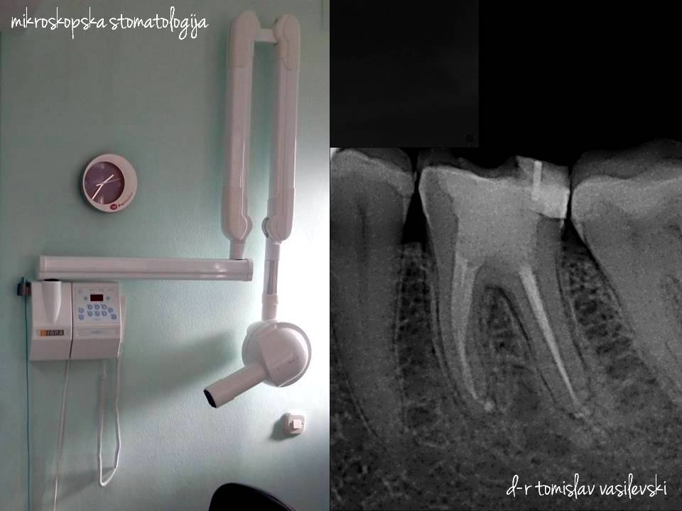
Latest microscopic root canal treatment
With contemporary methods of endodontic treatments, teeth can be saved in contrast to classical methods with which successfulness is less possible and are the main reason for removing of the teeth. Nothing can replace your natural tooth, even the latest technology making bridges and implants, therefore we need to do everything what is possible to safe it, before to decide to remove it.
Procedure of microscopic root canal treatment
1. Before starting with the treatment we are making digital x-ray record.

2. Then tooth is isolated with rubber dam which is made of rubber, ring which holds the rubber to the tooth, frame which holds the rubber, forceps for application, and forceps for perforating the rubber. This way is suitable for working in aseptic (sterile) conditions, and tooth is not in contact with bacteria from the mouth.

3. Helping with microscope, all canals can be found ( single tooth can have one, two, three, four and more canals). There are canals not visible to the naked eye, and if some of them are not found, treatment can not be successfully completed.

4. Lenth of the canals is precisely appoint first with digital apex locator (which measure precisely in all kind of conditions including dry, wet and canals with blood) and then with digital x-ray record, which is made with instruments in canals for even more precisely appoint of the canal length.

5. Mechanical processing of the canals to the tip of the root with endomotor and flexible expanders, with which easily are processed long, curved and narrowed canals in contrast to hand steel expanders.

6. Chemical processing of canals – heavily irrigation with solutions for disinfection which are heated on 60°C and are activated with endoactivator after application in the canal with what their efficiency in destroying bacterial is even better, not just in the main canal but also in the lateral canals.

7. Three dimensional filler of the canals with warm gutta percha which is melting on 200°C, and with what except main canals, are also filled lateral canals. By this way of filling, canals are hermetically closed and are impermeable for bacteria.

8. At the end is made one more digital x-ray record to ensure that canals are properly filled.

